
The Punishment Must Fit the Crime: A Kneecap Dislocation That Needed Two Surgeries
A kneecap dislocation with a torn MPFL, abnormal alignment and a cartilage injury, treated with MPFL reconstruction, a tibial tubercle osteotomy and MACI.
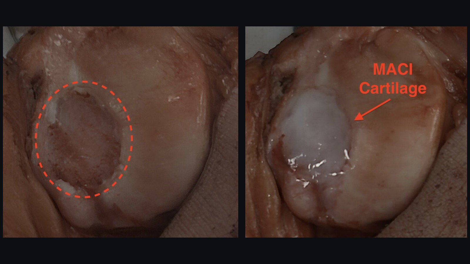
A man in his early 30s came to see me after his kneecap (patella) dislocated for the third time. The first dislocation was traumatic, when the side of a counter hit his knee. The second came after a slip and fall, and the third, which caused the cartilage injury, happened snowboarding. His goal is to get back to full activity, including snowboarding and basketball.
This case highlights my mentality on patellar dislocations: the punishment must fit the crime. Some patellar dislocations can be treated with physical therapy alone, and some require large operations. His was one of the large ones. He needed an MPFL reconstruction, a tibial tubercle osteotomy (TTO) and a MACI cartilage transplant.
Why a kneecap dislocates
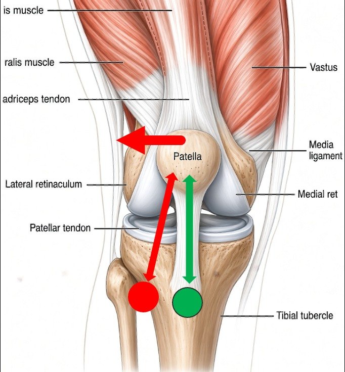
From an engineering perspective, the patella is at the whim of whatever is pulling on it. It is pulled by the quadriceps and the patellar tendon, contained by the groove at the end of the femur (the trochlea), and supported by the retinaculum and the MPFL. Depending on how all of these are tensioned and aligned, the patella can be pulled in different directions. All of its stability, or instability, comes from the forces these other structures put on it. That is why it is so critical to look at those structures to understand why the patella is dislocating.
What the imaging showed
After evaluation with X-ray and MRI, we found two reasons: he had torn his MPFL, and he has abnormal native alignment. We measure alignment on MRI with the TT-TG distance, which is how far the tibial tubercle sits from where the center of the patella should be tracking on the femur. We also take several other measurements, including patellar height, and we check the alignment of the hips for any rotation of the femur that makes the kneecap more likely to slide to the outside.
In addition, when he dislocated, he knocked off a large piece of cartilage from his patella, leaving a defect about 2 cm by 1.5 cm.
The plan: TTO, MPFL reconstruction and MACI
The tibial tubercle is the bump on the front of the shin bone (tibia) where the patellar tendon attaches. When it sits toward the outside, like the red line and circle, it puts a vector force on the patella that encourages it to dislocate. The solution here is to correct his alignment with a tibial tubercle osteotomy, moving the tubercle to the position of the green line and circle. This pulls the patella more in line with the quadriceps and allows for better patellar tracking.
Pretty much every kneecap dislocation tears the MPFL. The MPFL is the stabilizing ligament on the inner side of the knee, so when the kneecap dislocates far enough to the outside, that ligament tears. When a patellar dislocation is treated surgically, an MPFL reconstruction is almost always part of the operation. We rebuild it with a graft that is actually stronger than the native ligament, so it is a pretty powerful restraint against another dislocation.
Because of the cartilage injury, he also needed a cartilage transplantation with MACI. The other common option for cartilage restoration is an osteochondral allograft, but on the patella that is a challenge. The kneecap has unusual contours and it can be hard to get that graft to heal, so for the patella, MACI is preferred.
The two surgeries
MACI requires two operations. The first is an arthroscopy, where we removed the loose cartilage fragment and took a small biopsy of healthy cartilage. The biopsy goes to a lab near Boston that grows a sheet of his own cartilage cells, which takes about four to six weeks. That sheet is then transplanted back into the knee, where it matures into his own cartilage.
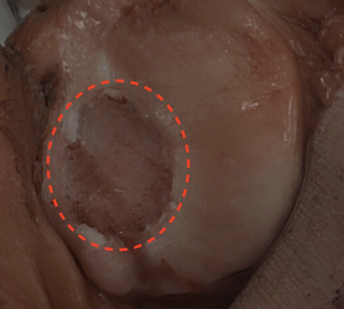
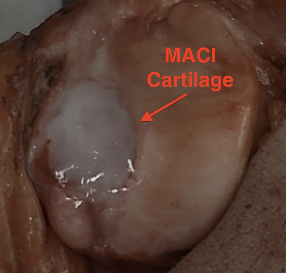
Everything else was done at the second surgery: the MPFL reconstruction, the tibial tubercle osteotomy, the MACI cartilage transplantation, and a lateral retinacular lengthening. It all happens at the second operation because that is the surgery where we actually open up the knee.
Recovery
He is freshly post-op, so I do not know about his full return yet. For the first 6 weeks he is toe-touch weight-bearing, but he can start range of motion right away. Full recovery is likely 9 months to a year, to give the osteotomy and the cartilage time to heal.
Who this applies to
The big highlight of this case is that no two patients are alike. With patellar dislocations especially, we need to look at the patient's alignment, their history of dislocations or injuries, associated injuries like an MPFL tear or cartilage damage, and their overall goals.
Our thinking about patellar dislocations has really changed. When I started my career, the common saying was “three strikes and you're done,” meaning we did nothing until someone had dislocated three times. Now we can better predict who is going to keep dislocating by looking at their anatomy and their exam. If someone is at very high risk of dislocating again, it is best to treat them early. That can mean a short course of physical therapy to see how they do, or in some cases going straight to surgery if there is cartilage damage or other damage that needs to be addressed. There are still lots of people who can be treated with physical therapy and bracing alone, if their anatomy is low risk and it is their first dislocation.
This article is general education, not medical advice for your situation. Every knee and hip is different — talk with your own doctor about your care.